Tissue Diagram Labelled
What is nervous tissue ? draw a well labelled diagram of neuron Draw a labelled diagram of xylem tissues Connective physiology tissues epithelial fishersci danlos ehlers different dense cartilage mcqs textbook eds histology areolar cells organs supporting elastic syndromes
Draw a labelled diagram of xylem tissues - 1n3ed4yy
Anatomy tissue quizlet With help of neat labelled diagram, describe the structure of areolar Xylem diagram labelled tissues draw topperlearning biology kolte sheetal answered jul 2nd pm
Histology image: connective tissue
Nerve cell human body tissues stock illustration different types cells depositphotos kinds som intro histology siu blueringmediaTissue connective tissues types bone functions body diagram cells human areolar blood loose anatomy cartilage cell matrix labeled animal physiology Tissues basic ppt chapter body cover powerpoint presentation surfaces glands hollow epithelial ducts cavities organs form line2-3 tissues.
Tissues histology quizletMuscle tissue drawing at paintingvalley.com Unit 6: tissue structure and functions – douglas college human anatomyTissue anatomy and physiology.
Anatomy physiology tissue chapter tissues types human body cells study cell biology slideshare systems
Physiology tissueDraw a labelled diagram showing location of different types of Tissue connective histology components features generalMuscle tissue drawing skeletal types muscular anatomy cell biology smooth cells human body cardiac diagram tissues functions muscles histology fibers.
Areolar connective diagram labelled tissue structure neat cells describe tissues help reticularAnatomy physiology tissue Tissues body cells tissue biology anatomy human physiology types different google science histology nursing school study connective identify major cellTissue tissues anatomy epithelial nervous physiology.

Epithelium cells histology layer nuclei membranous columnar stratified pseudo single two variable shape level height
Anatomy and physiology tissueAnatomy and physiology tissue chapter Histology image: membranous epitheliumNeuron tissue nervous diagram labelled draw cell well parts.
Bone connective tissue diagramAnatomy and physiology tissues 😎 different kinds of tissues in the human body. types of tissuesLocation tissue meristematic diagram types labelled draw different showing meristem lateral apical biology primary.


what is nervous tissue ? draw a well labelled diagram of neuron

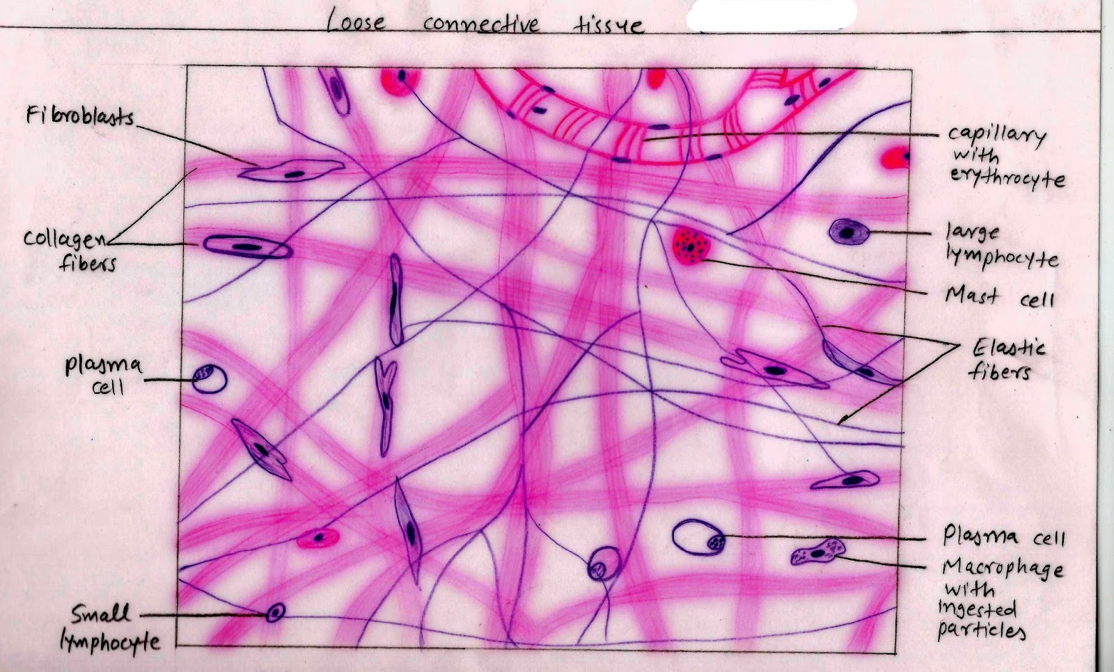
with help of neat labelled diagram, describe the structure of areolar

Muscle Tissue Drawing at PaintingValley.com | Explore collection of

Unit 6: Tissue Structure and Functions – Douglas College Human Anatomy

Anatomy And Physiology Tissue

😎 Different kinds of tissues in the human body. Types of Tissues

Bone Connective Tissue Diagram

2-3 Tissues - Anatomy & Physiology

Anatomy And Physiology Tissues